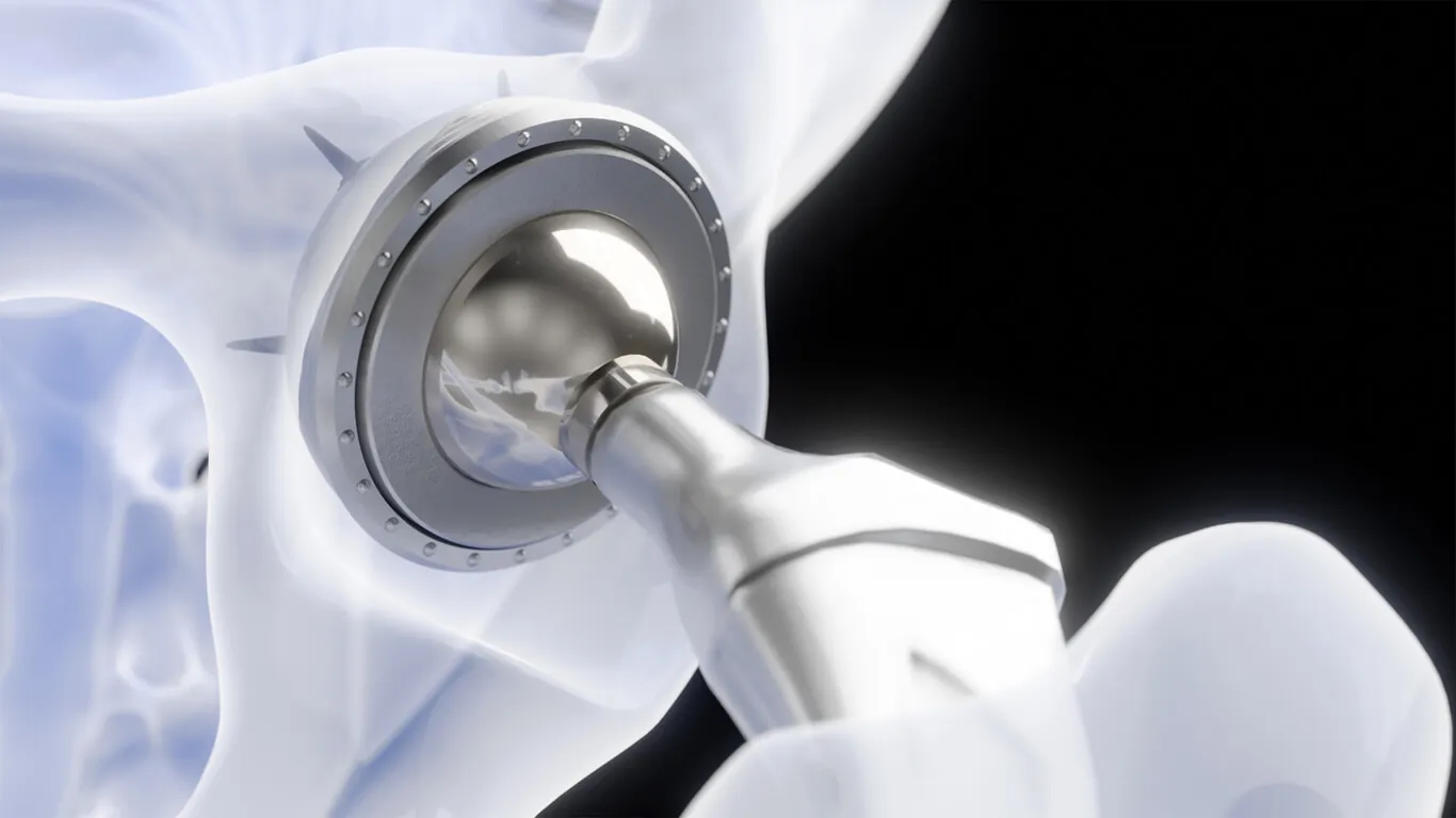
Avascular necrosis (AVN) of the hip — also called osteonecrosis — is the death of bone in the femoral head caused by loss of its blood supply. Dr. Vinay S. Joshi treats AVN at Kokilaben Dhirubhai Ambani Hospital, Mumbai, offering joint-preserving Regrow therapy for early-stage disease and ceramic-on-ceramic hip replacement once the femoral head has collapsed.
AVN behaves differently from ordinary arthritis in one decisive respect: it is a race against structural collapse. Before the femoral head loses its shape, the hip can often be saved. After it collapses, replacement becomes the only reliable option. Everything about AVN management follows from where the patient sits on that line — which is why an early MRI is the most consequential step in the whole pathway.
Why AVN matters particularly in India
AVN of the hip is one of the commonest reasons for hip replacement in younger patients in India, frequently presenting in people under 50 and sometimes in their twenties and thirties. Recognised associations include high-dose or prolonged corticosteroid therapy, significant alcohol intake, sickle cell disease, and trauma such as a femoral neck fracture or hip dislocation. A proportion of cases are idiopathic, with no identifiable cause.
The age profile is what makes AVN clinically demanding. A hip replacement in a 35-year-old must last decades, which shapes every decision about bearing surface, fixation and — where possible — whether replacement can be avoided at all for now.
The stages of AVN
Staging determines treatment, and the pivotal distinction is whether the femoral head has collapsed.
| Stage | What is happening | Typical treatment |
|---|---|---|
| Stage I | Bone tissue is dying. X-rays are normal; only MRI detects it. The femoral head is fully spherical. | Joint preservation — core decompression, with biological augmentation |
| Stage II | Bone changes visible on X-ray as sclerosis or cysts. The head remains spherical. | Joint preservation, best results in stage IIA with a small lesion |
| Stage III | Subchondral fracture — the 'crescent sign'. The head has begun to collapse. | Hip replacement in most cases |
| Stage IV | The head is deformed and secondary arthritis has developed in the joint. | Total hip replacement |
In stages I and II an X-ray is frequently normal. A patient with unexplained groin or hip pain, particularly with any AVN risk factor, needs an MRI — not reassurance from a clear radiograph. The window in which the hip can be preserved is measured in months, and it closes silently.
Symptoms
- Groin pain, sometimes referred to the buttock, thigh or knee — knee pain as the presenting symptom of hip disease is common and frequently misdirects investigation.
- Pain that is worse on weight-bearing and eases with rest, at least initially.
- Progressive restriction of hip movement, particularly internal rotation.
- Night pain and pain at rest as the disease advances.
- A limp, and in later stages a shortened leg as the head collapses.
Core decompression for early AVN
In stages I and II, Dr. Joshi performs core decompression: a precise channel is drilled under fluoroscopic guidance from the lateral femur, through the neck, into the necrotic segment. This relieves the raised pressure inside the bone, which is itself part of what perpetuates the ischaemia, and opens a route for new blood vessels to grow into the dead zone.
On its own, core decompression halts progression in a meaningful proportion of early cases. Its results improve when the decompressed channel is biologically augmented — which is the basis of Regrow therapy.
Regrow therapy: preserving the joint
Regrow therapy combines core decompression with cell-based and biological augmentation, with the aim of regenerating bone and restoring blood supply rather than replacing the joint.
What the procedure involves
- Bone marrow is aspirated from the patient's own iliac crest.
- It is processed at the bedside to concentrate the mesenchymal stem cells and progenitor cells it contains.
- Core decompression is performed under fluoroscopic guidance into the necrotic segment.
- The concentrated marrow aspirate is delivered into the decompressed zone, together with platelet-rich plasma and bone graft material.
- These stimulate new bone formation and vascular ingrowth, repairing the segment from within.
Who it suits
The best candidates are patients with stage I or stage IIA disease, in whom the femoral head is still spherical and intact, typically under 50, with a necrotic lesion occupying less than about 30% of the head. Lesion size and location matter as much as stage — a large lesion under the weight-bearing dome carries a worse prognosis than a small medial one.
Results and follow-up
In appropriately selected patients, the published literature reports arrest of progression in roughly 70–80% of cases, with many patients experiencing significant pain relief within three to six months. Those are figures from published series rather than from Dr. Joshi's own practice — he does not hold separate outcome data for this procedure, and prefers to quote the evidence base rather than an in-house number. Serial MRI at six and twelve months monitors healing. If the disease progresses despite treatment, hip replacement remains fully available — the joint-preserving attempt does not compromise a later replacement.
Hip replacement for collapsed AVN
Once the femoral head has collapsed — stage III or IV — replacement becomes necessary. AVN replacement is technically distinct from replacement for ordinary osteoarthritis in three ways: the bone geometry is abnormal, there is bone deficiency in the necrotic zone, and the patient is usually young enough to need exceptional implant longevity.
Dr. Joshi typically uses uncemented fixation with a ceramic-on-ceramic bearing for AVN patients. The reasoning is durability: ceramic-on-ceramic produces up to 4,000 times less wear debris than metal-on-polyethylene, and wear debris is what ultimately loosens a hip. For a patient who needs a 25 to 30 year implant life, that difference is decisive. Costs for each bearing option are set out in the hip replacement cost guide.
If both hips are affected
Bilateral AVN is common, particularly in steroid-associated and sickle-cell-associated disease. It is not unusual for one hip to be collapsed while the other is still at stage I or II — in which case the two hips need entirely different treatments, and the second hip may be saveable if it is investigated rather than assumed to be following the first. Dr. Joshi images both hips whenever AVN is diagnosed on one side.
Frequently Asked Questions

Consultant Orthopaedic & Joint Replacement Surgeon and lead of the Arthroplasty team at Kokilaben Dhirubhai Ambani Hospital, Mumbai. Twelve years of specialist training across five UK orthopaedic centres, with further fellowships in Canada, Italy, Singapore and the USA. More than 4,500 joint replacements performed.